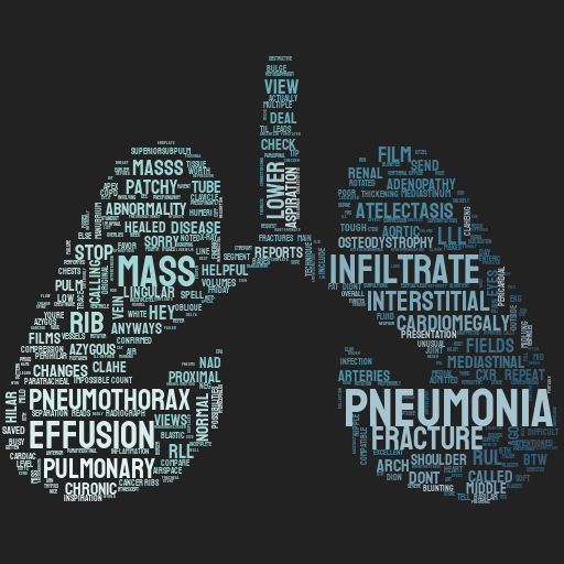
(opacity proportionate to frequency)
Hover for descriptions. Click for examples.
ChestSearch
- Masses/nodules can be subtle areas of increased density, sometimes seen under bones, or a deformity of mediastinal contours
- Fractures can be subtle. Look carefully at the ribs, clavicle, spine and scapulae for discontinuities or cortical step-off. History and location of pain can be helpful
- Check the costophrenic angles for small effusions
- For PTX, look for increased lucency in the chest, loss of peripheral lung markings or a visible pleural edge
- Pulmonary edema clues include: upper lobe pulmonary venous enlargement, cardiomegaly, peribronchial cuffing, Kerley lines, bilateral perihilar opacities and pleural effusions.
- Adenopathy can show abnormal enlargement/fullness of the hila or superior mediastinum
- Subtle enlargement of the cardiac silhouette can be detected by measuring the cardiothoracic ratio
- Subdiaphragmatic free air or visualizing the diaphragm continuously across the midline are signs of pneumoperitoneum
- Pneumonia and other opacities can be subtle areas of increased density, sometimes obscured by the heart, ribs or other structures. Laterals can be helpful


