(diameter proportionate to frequency)
Hover for descriptions. Click for examples.

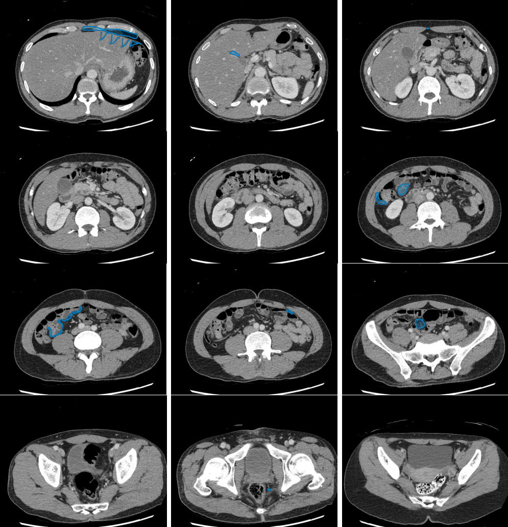
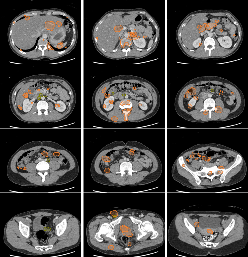
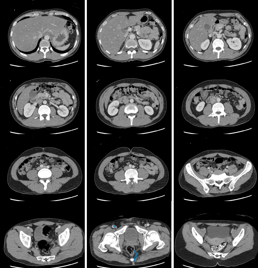
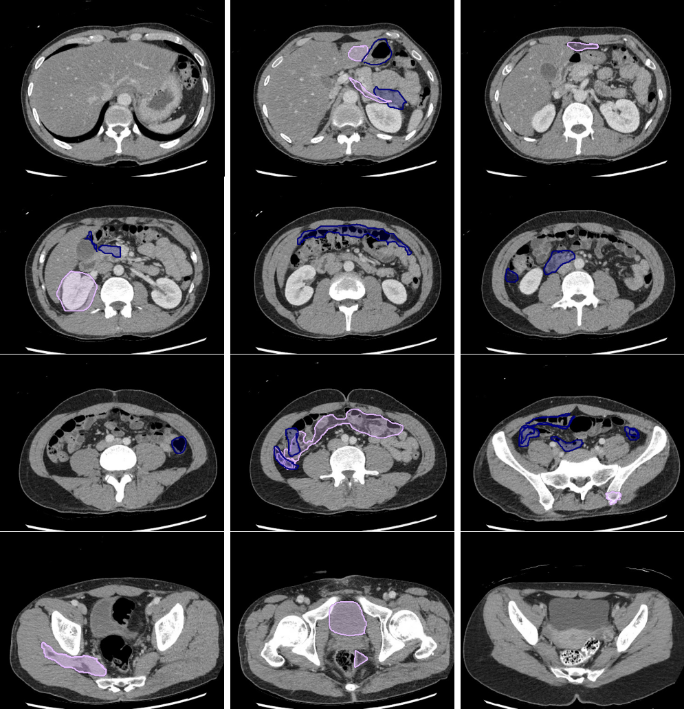
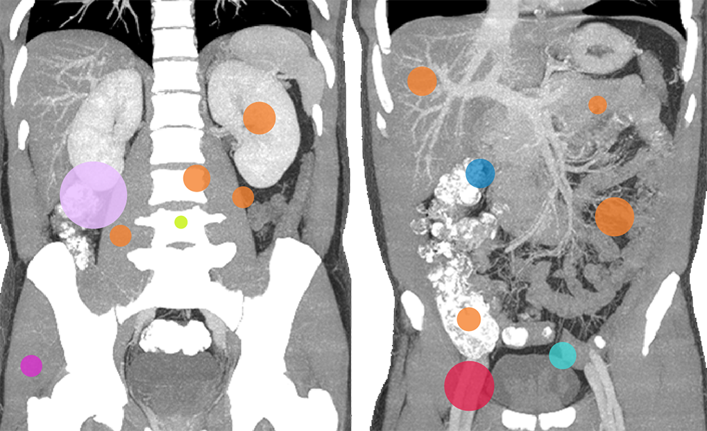
Body Search
- Liver lesions can be subtle. Use a "Liver" window to increase sensitivity.
- Review tumor markers.
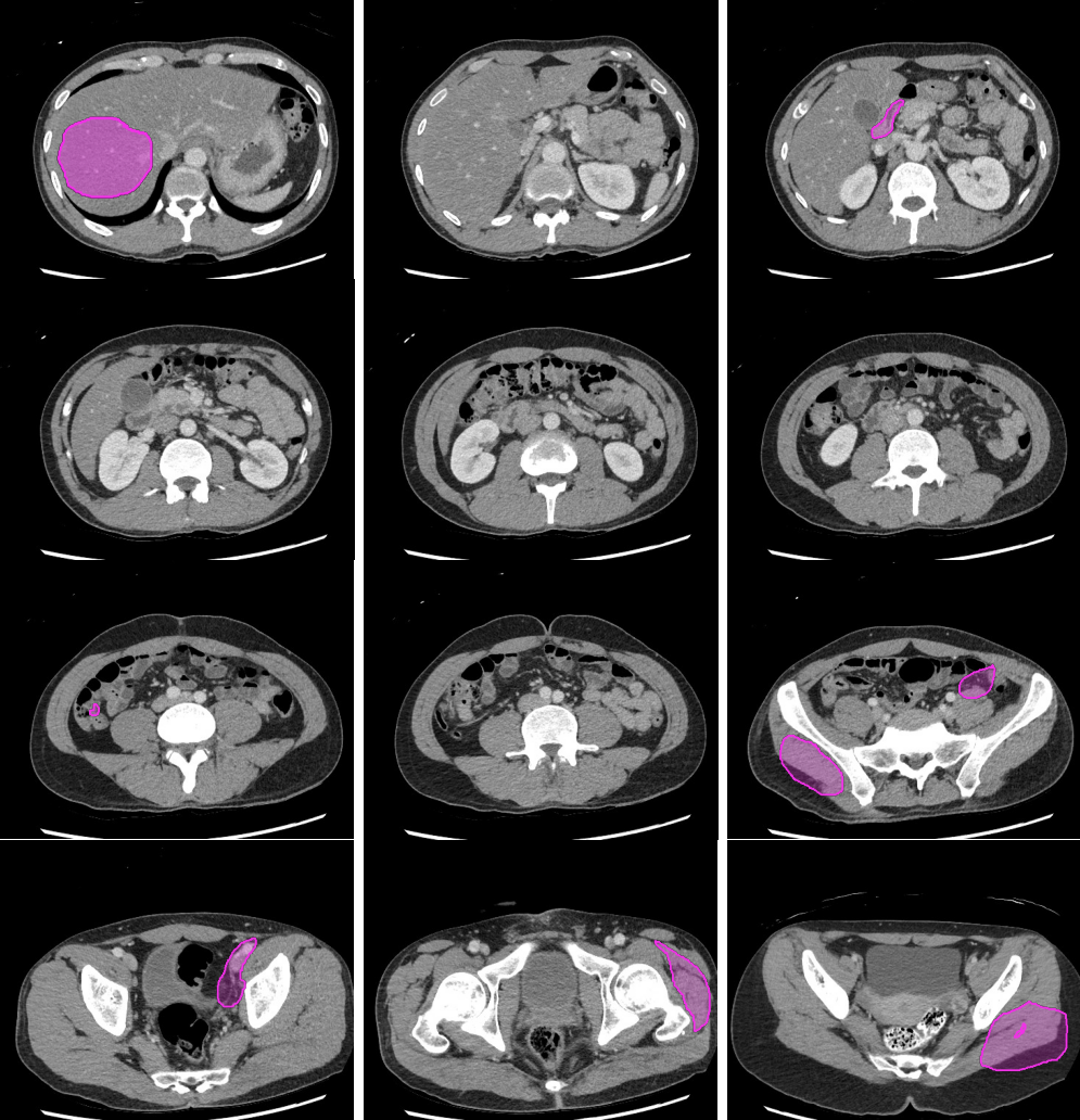
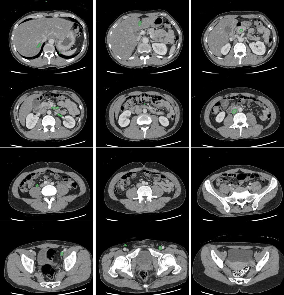
- Masses in the mesentery/peritoneum are often overlooked. Take advantage of multiplanar imaging and look in characteristic locations for peritoneal lesions.
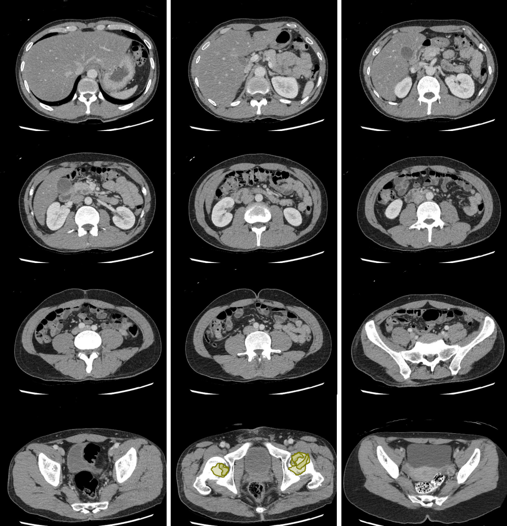
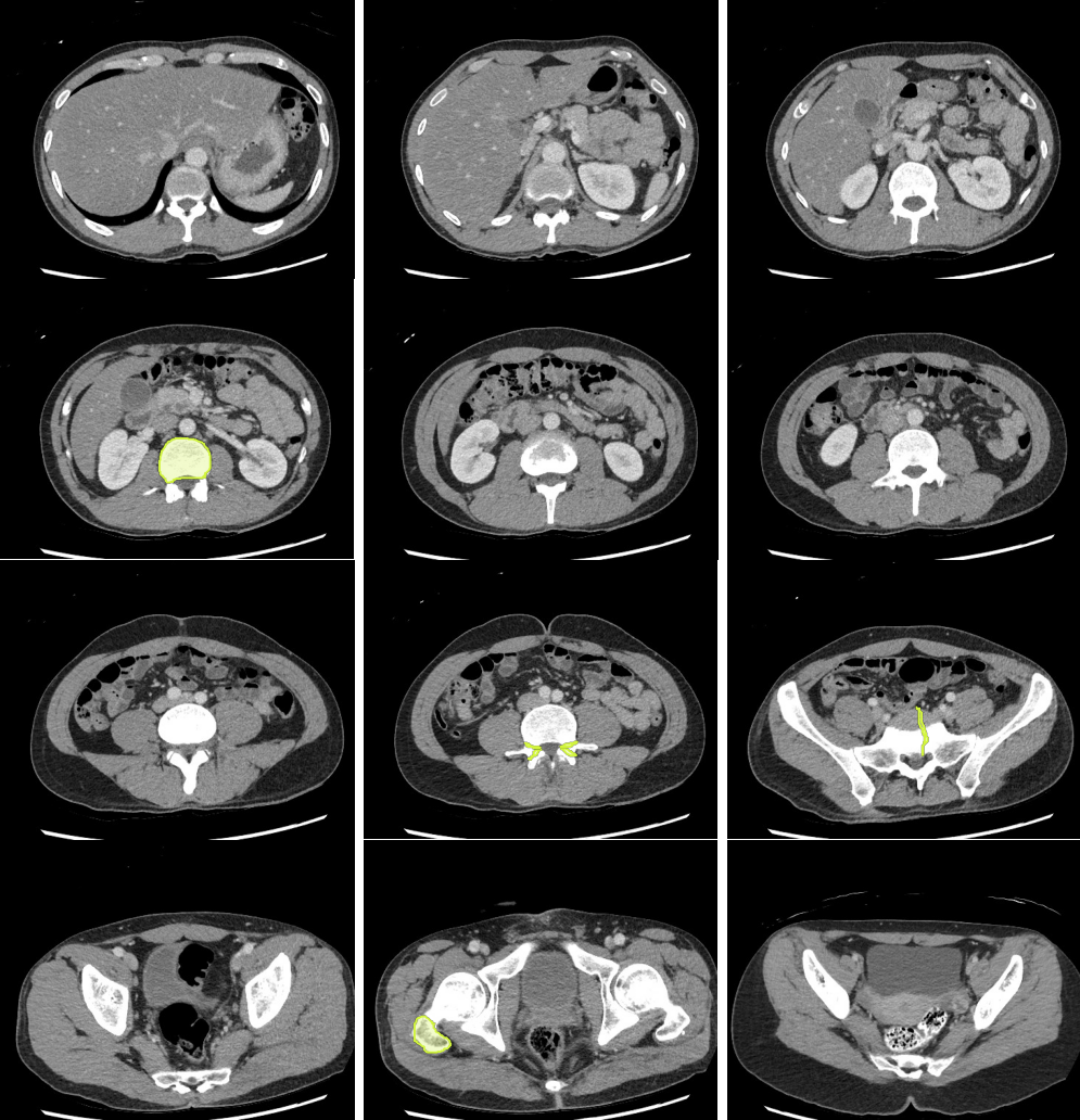
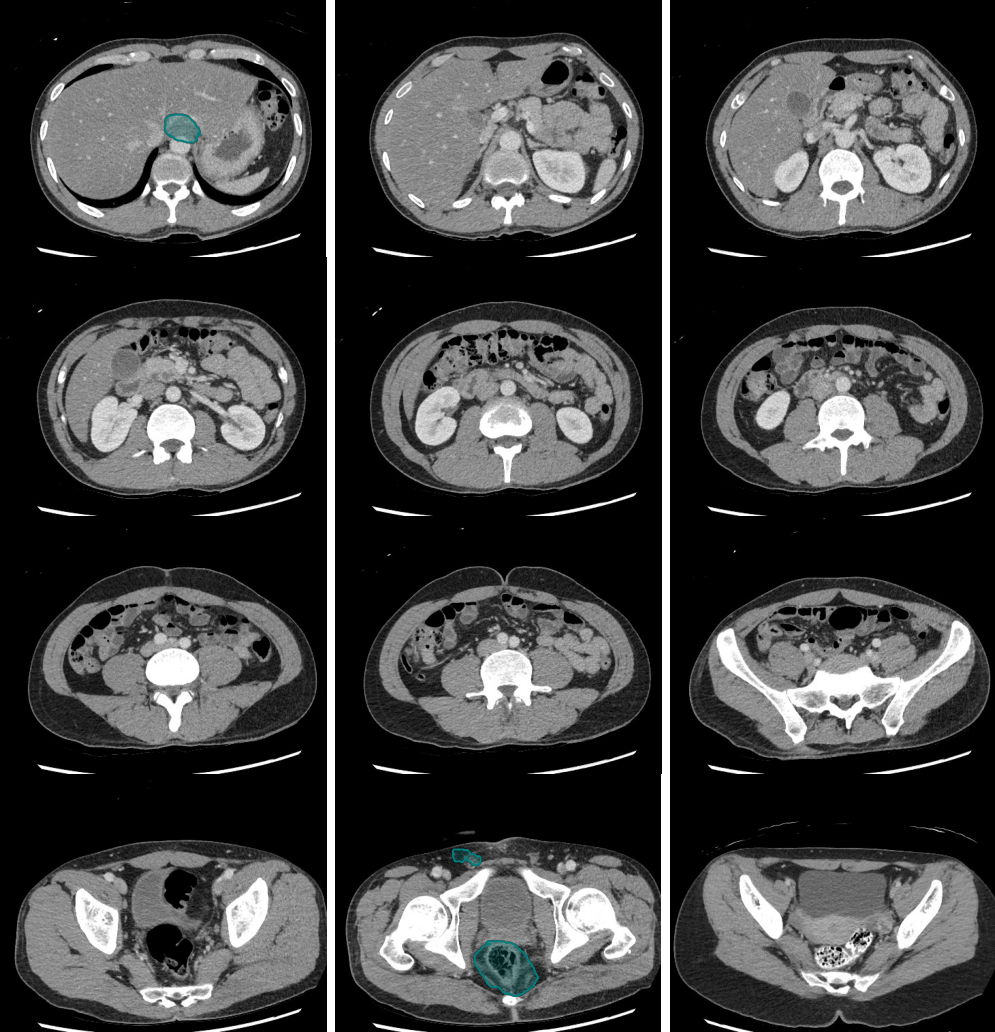
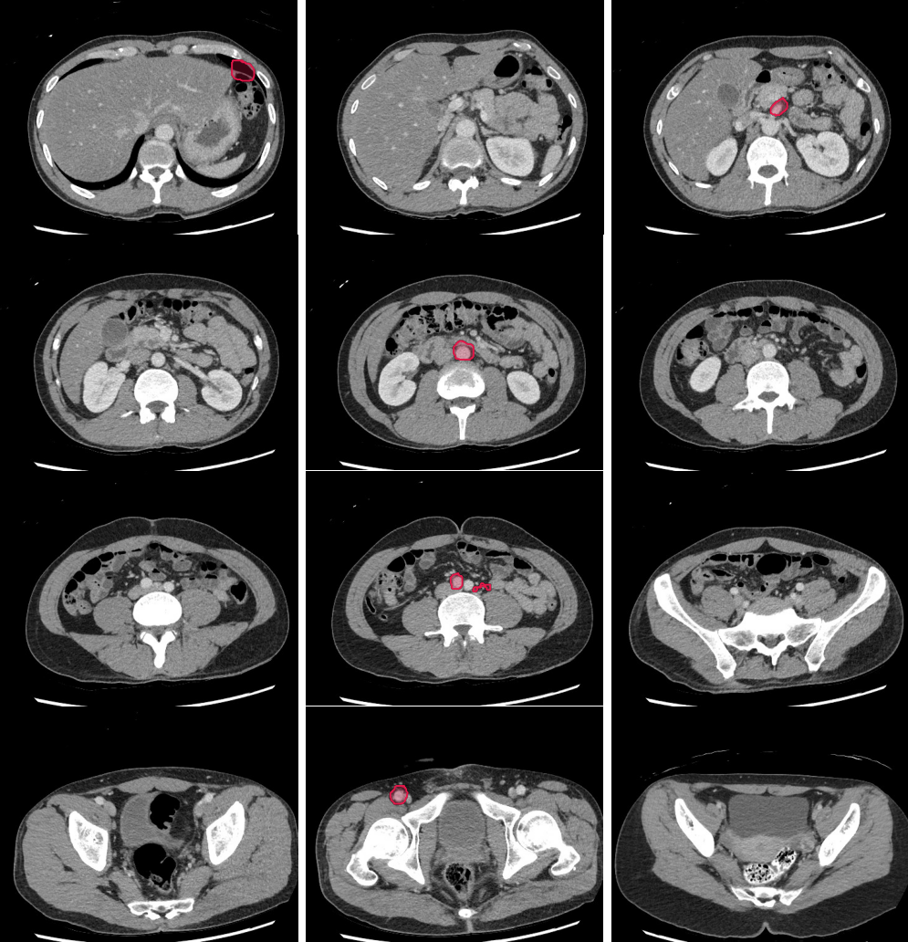
- Kidney masses are also overlooked. Look carefully at the collecting system and for renal contour abnormalities.
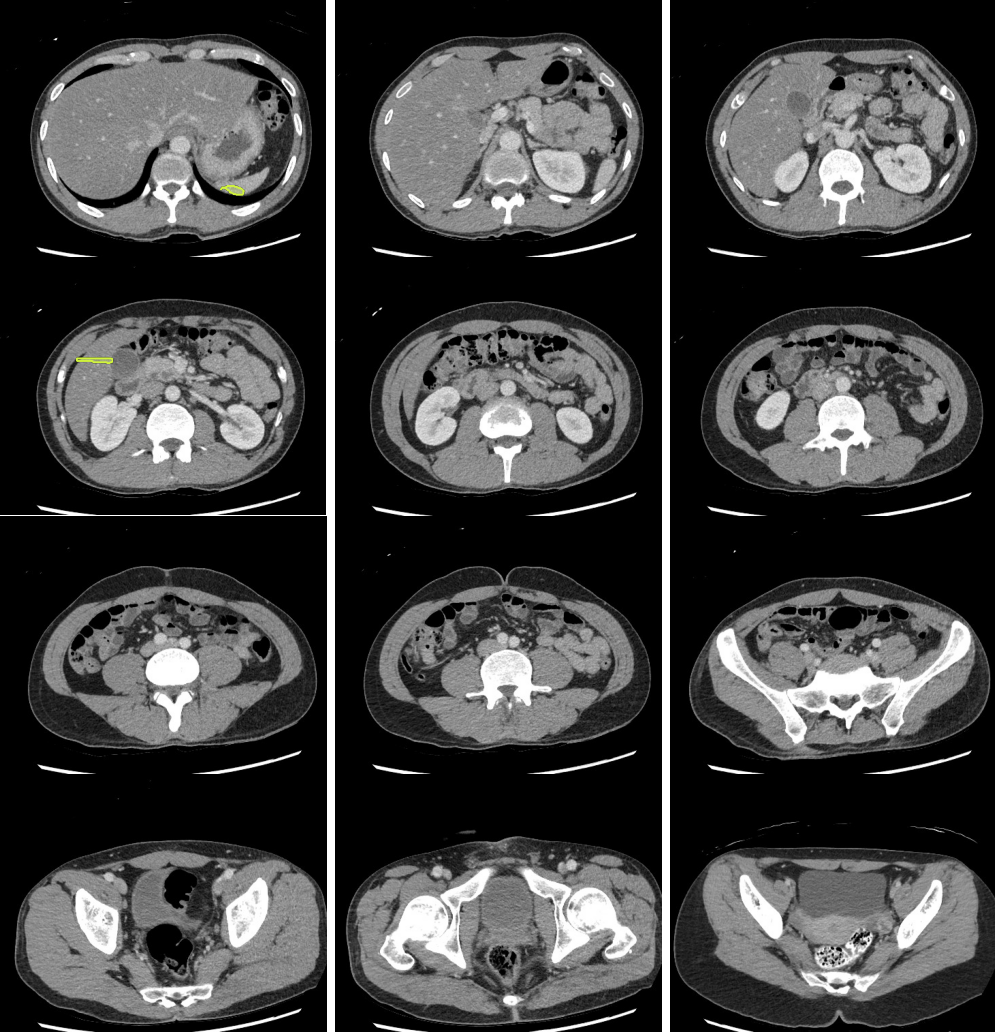
- Do not forget the bones. Pay attention to endplates and cortical margins.
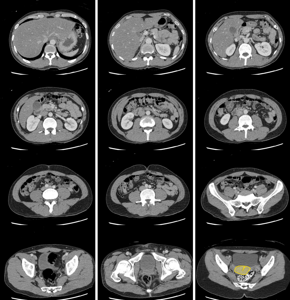
- Do not forget the paraspinal soft tissues. Using narrow windows to detect soft tissue abnormalities is useful.
- “Running the bowel” or tracking from above from the esophagus through the duodenal sweep and into at least the proximal jejunum and from below through the ileocecal valve into the terminal ileum is critical.
- Watch for wall edema and surrounding fatty infiltration as a sign of infection/inflammation associated with the digestive tract, especially the appendix.
- Look for filling defects in the vessels. Venous enlargement might indicate acute thrombus and confirming increased density by applying tight windows to potentially detect acute venous thrombus can be useful.
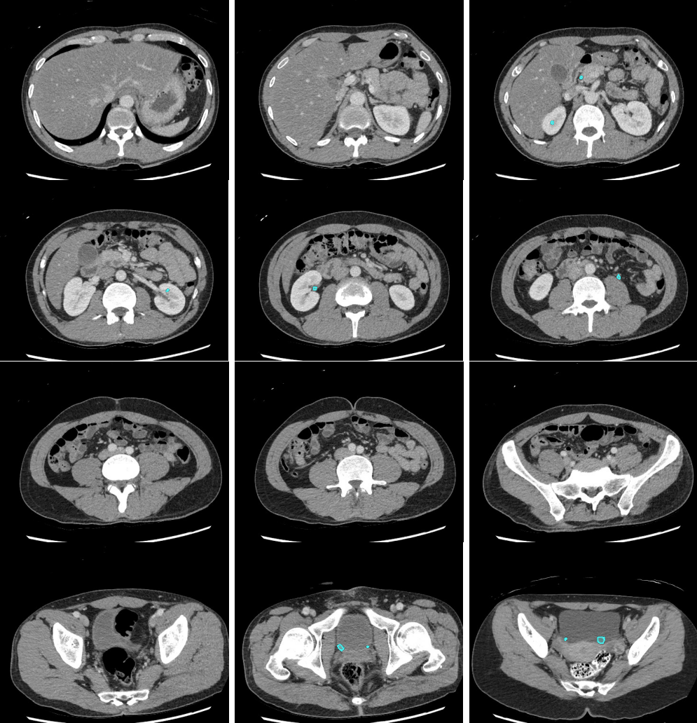
- Even without hydronephrosis, consider the possibility of ureteral calculi and attempt to follow the course of the ureters to screen for stones.
- Look carefully for small stones in the ureter/UVJ. Familiarize yourself with the normal location of the ureterovesical junctions and the configuration of the distal ureter to discriminate phleboliths from potential distal ureteral calculi